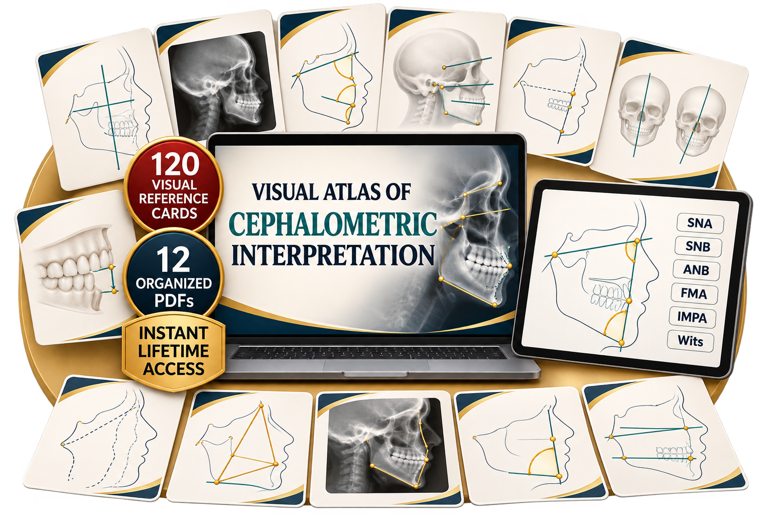
120 visual reference cards to
turn measurements and tracings
into clear clinical interpretation
Understand landmarks, planes, angles, skeletal patterns, growth, dental position, and soft tissues through a practical visual system that is easy to reference.
- 100% digital material
- Instant access
- Study on any device
- Secure payment
See what’s included
Explore selected cards from the Visual System for Cephalometric Interpretation.
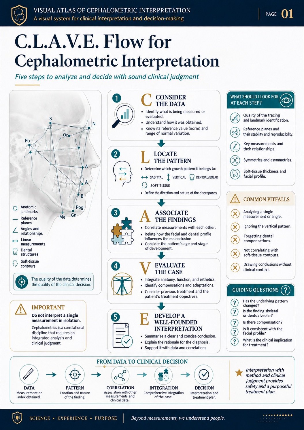
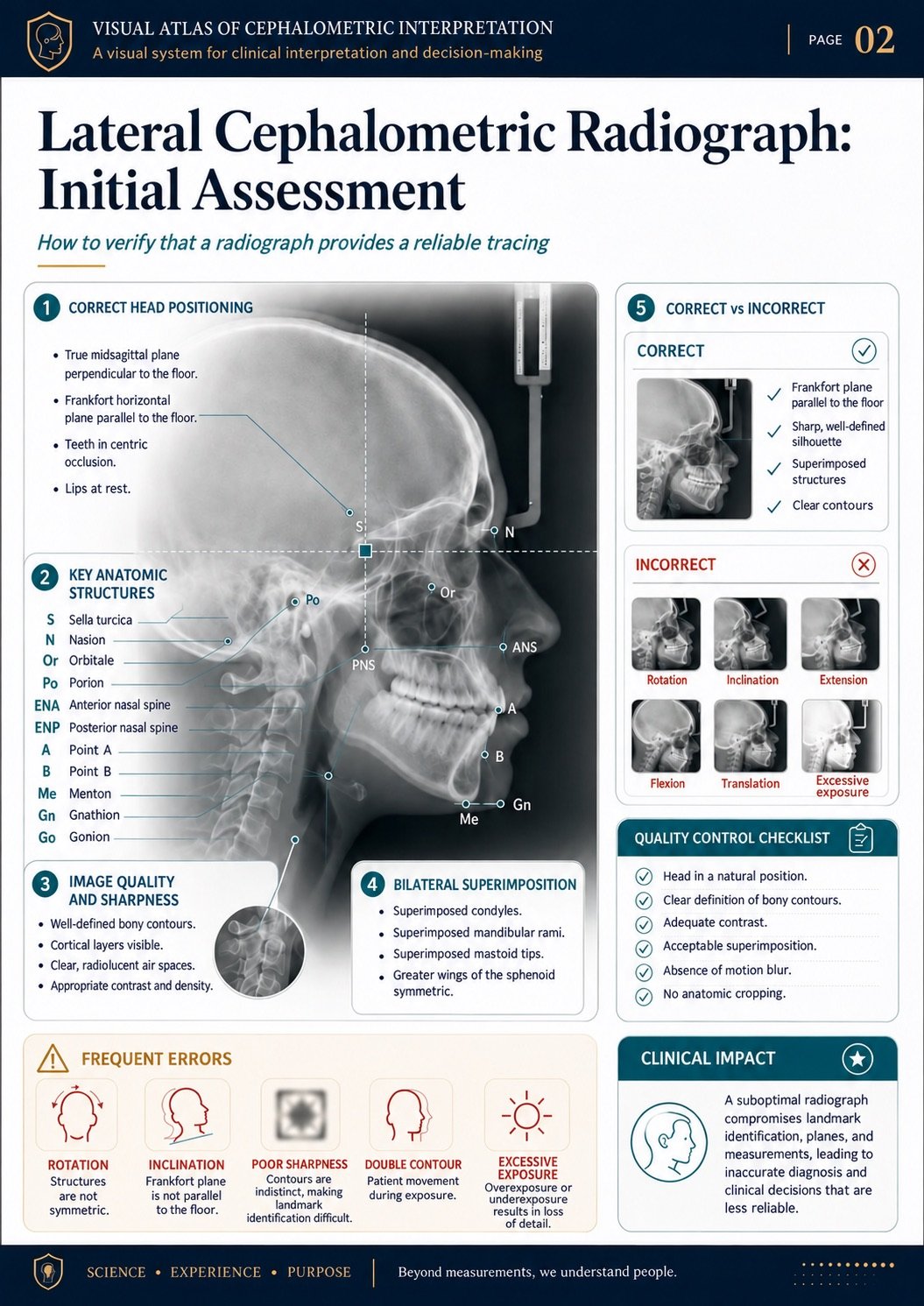
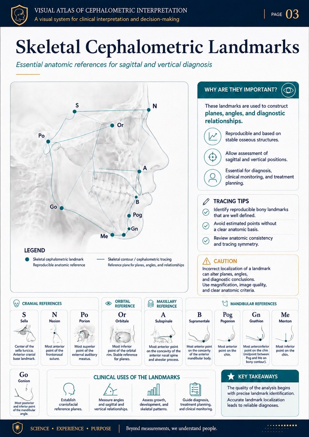
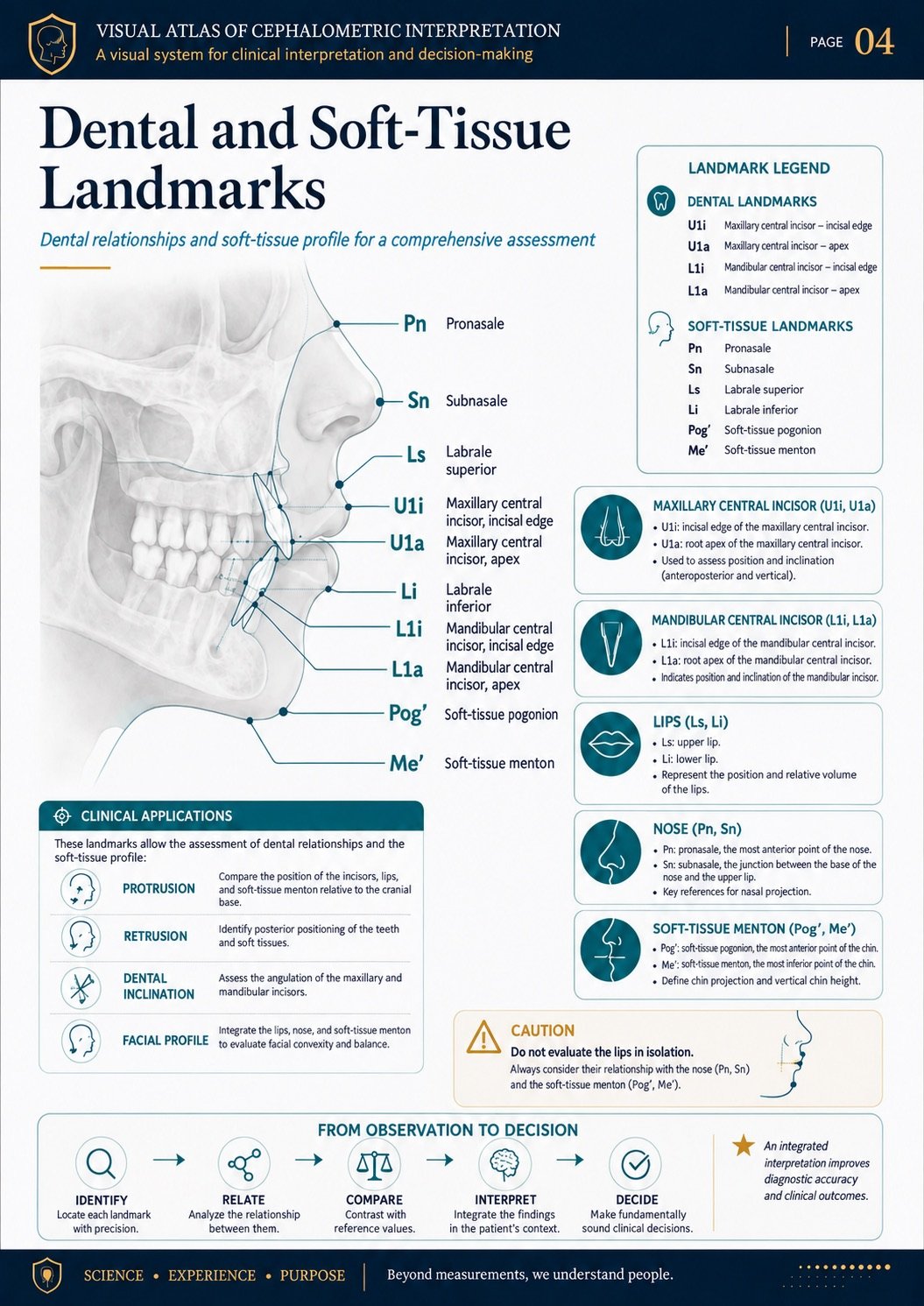
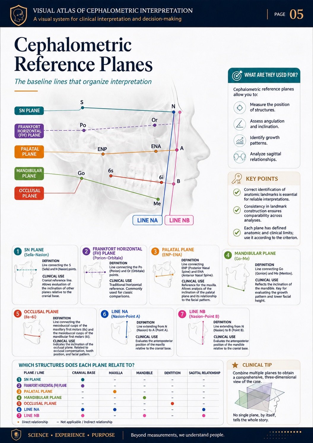
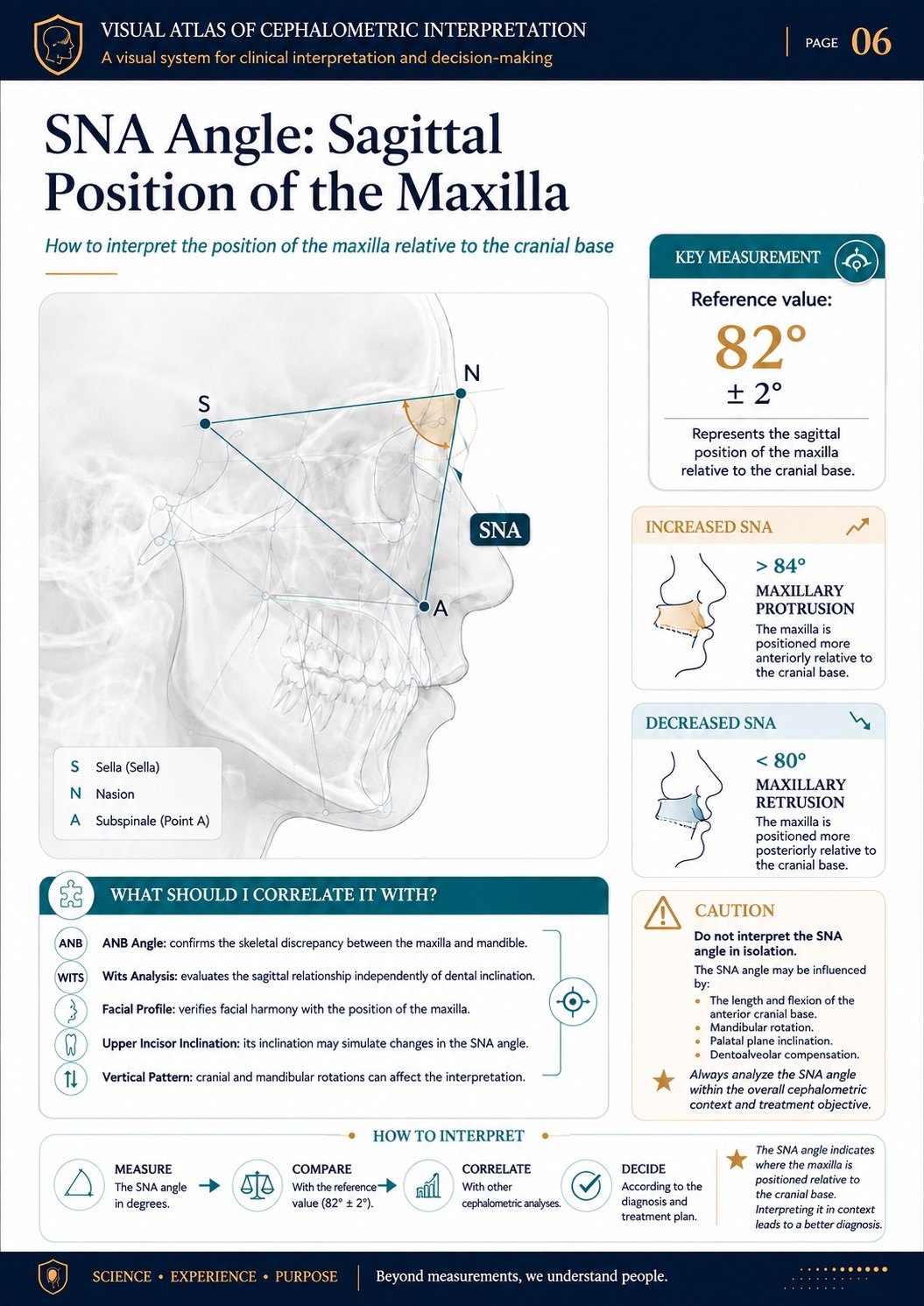
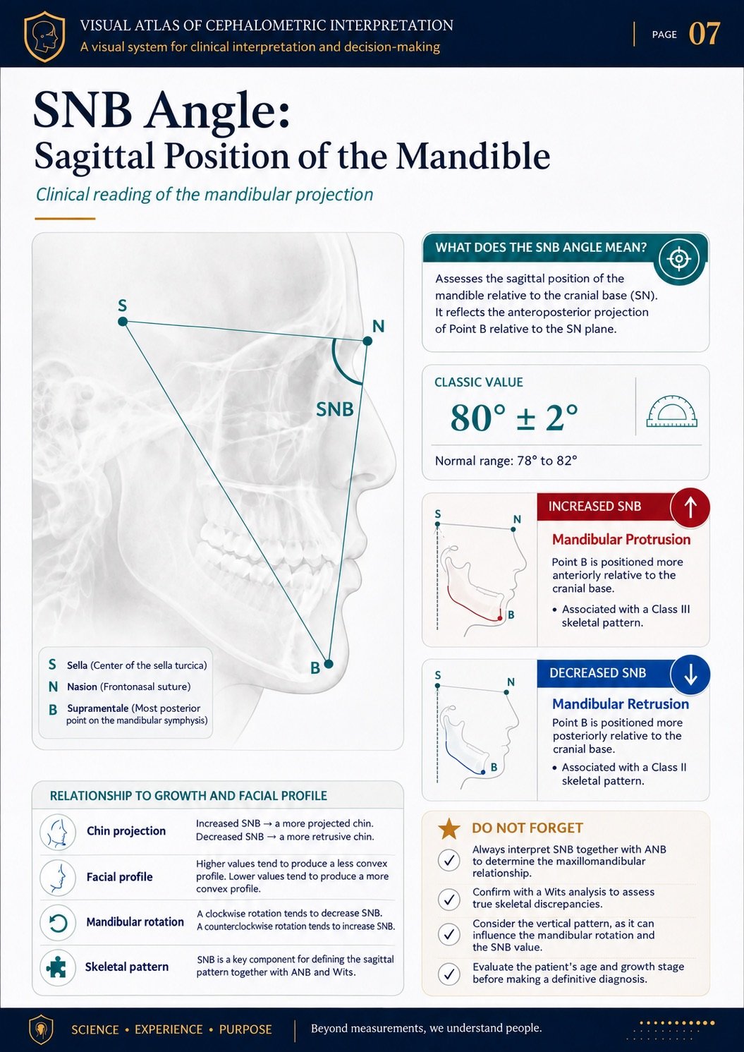
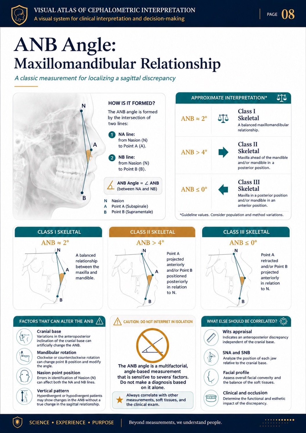
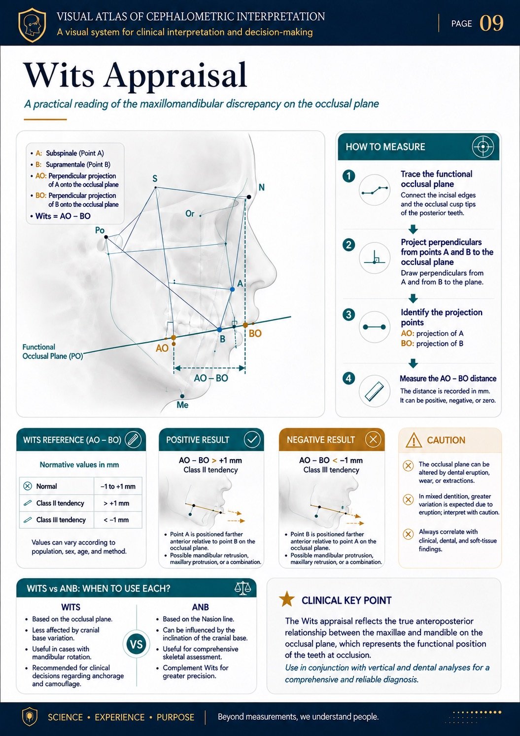
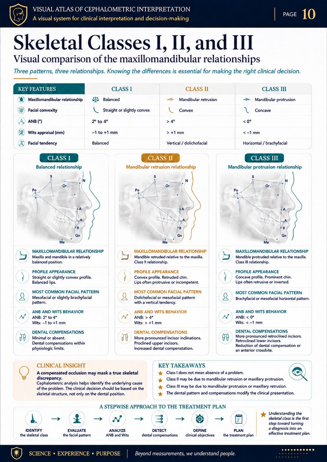
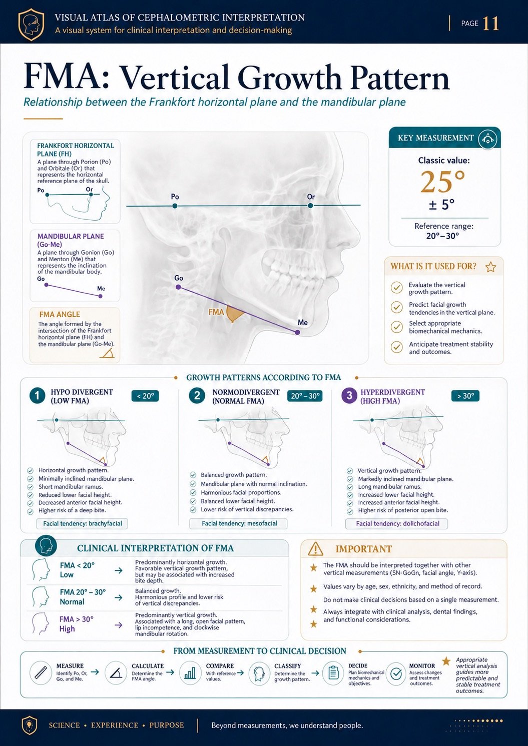
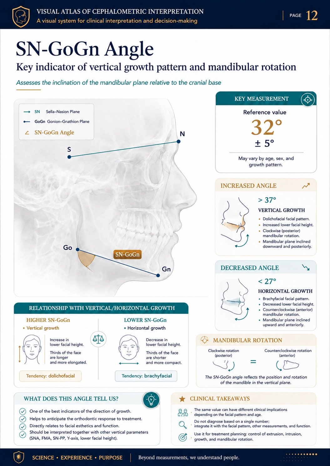
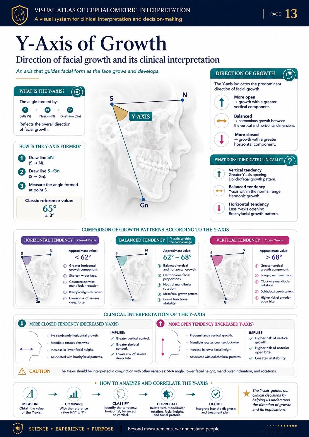
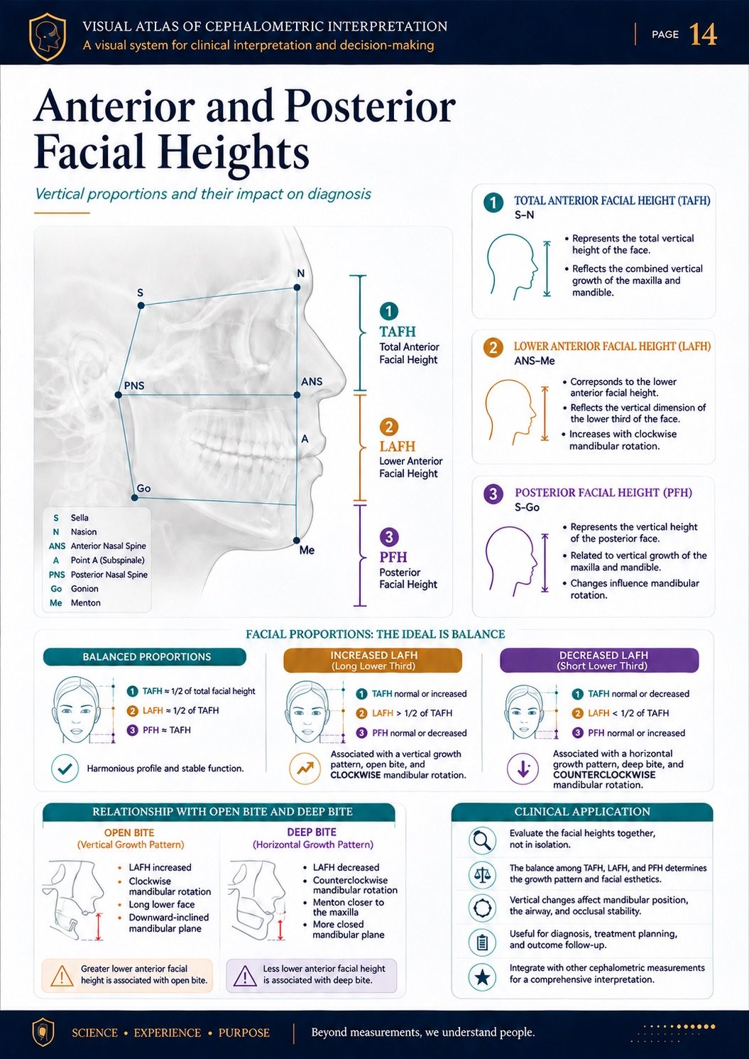
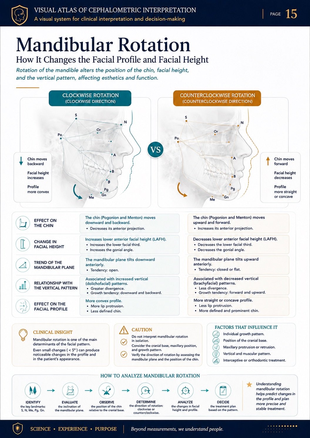
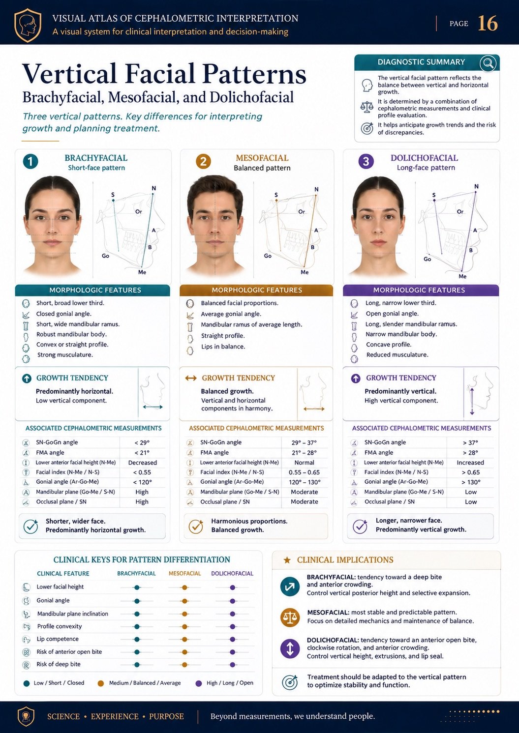
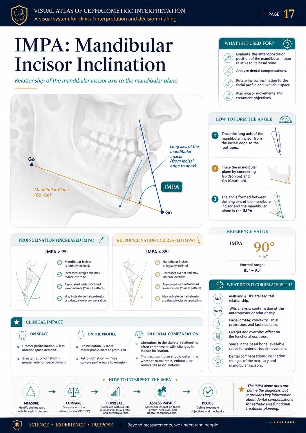
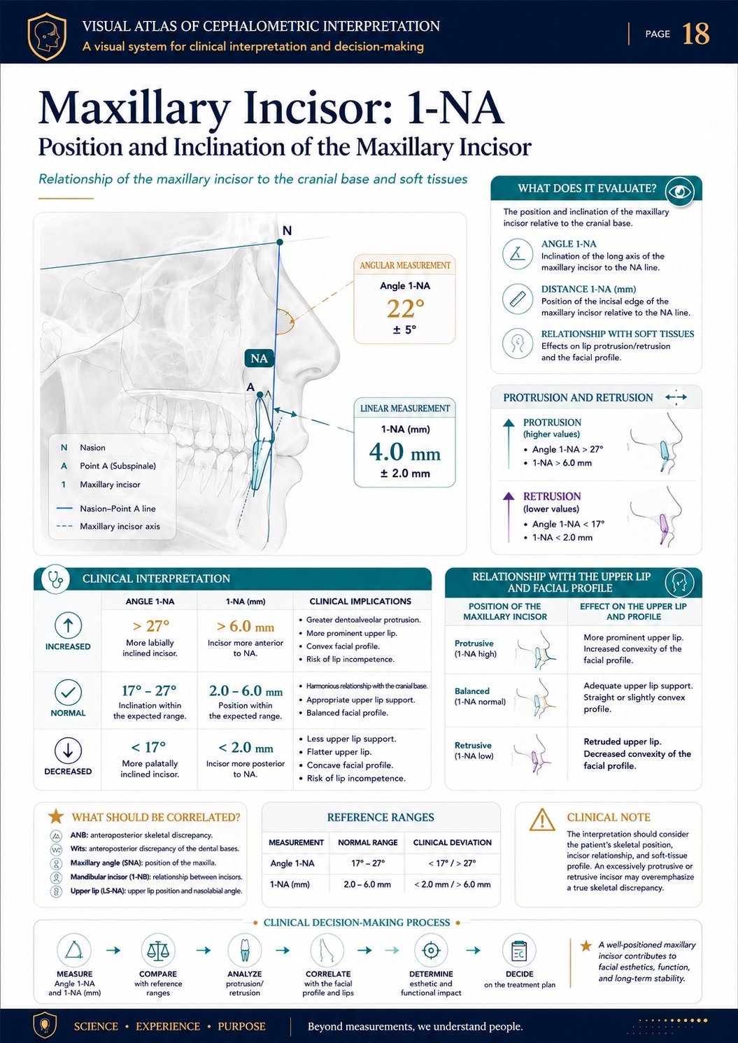
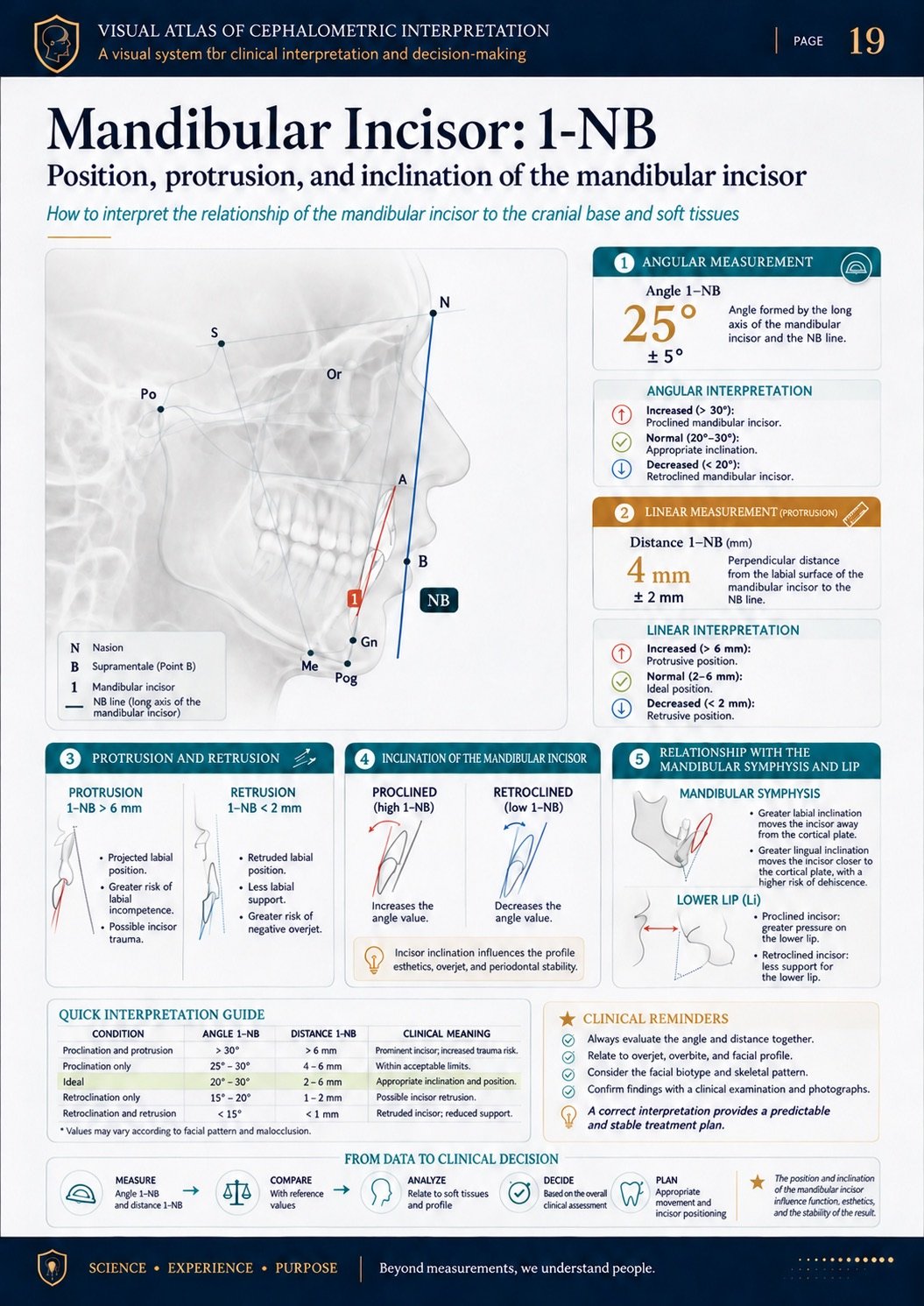
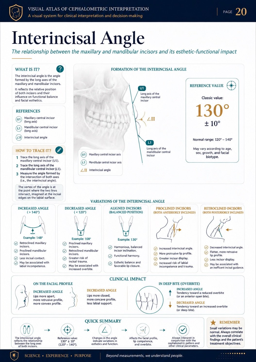
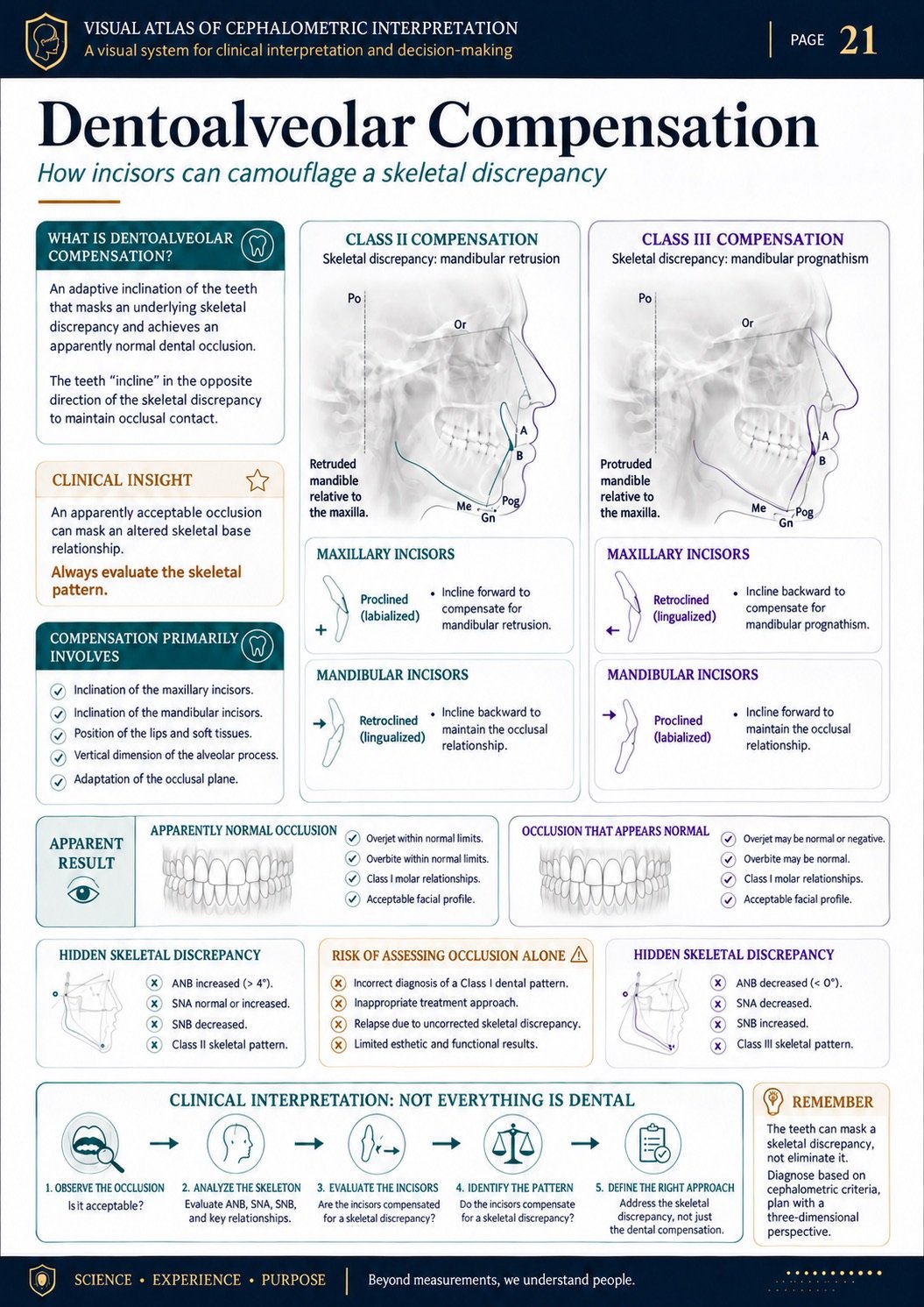
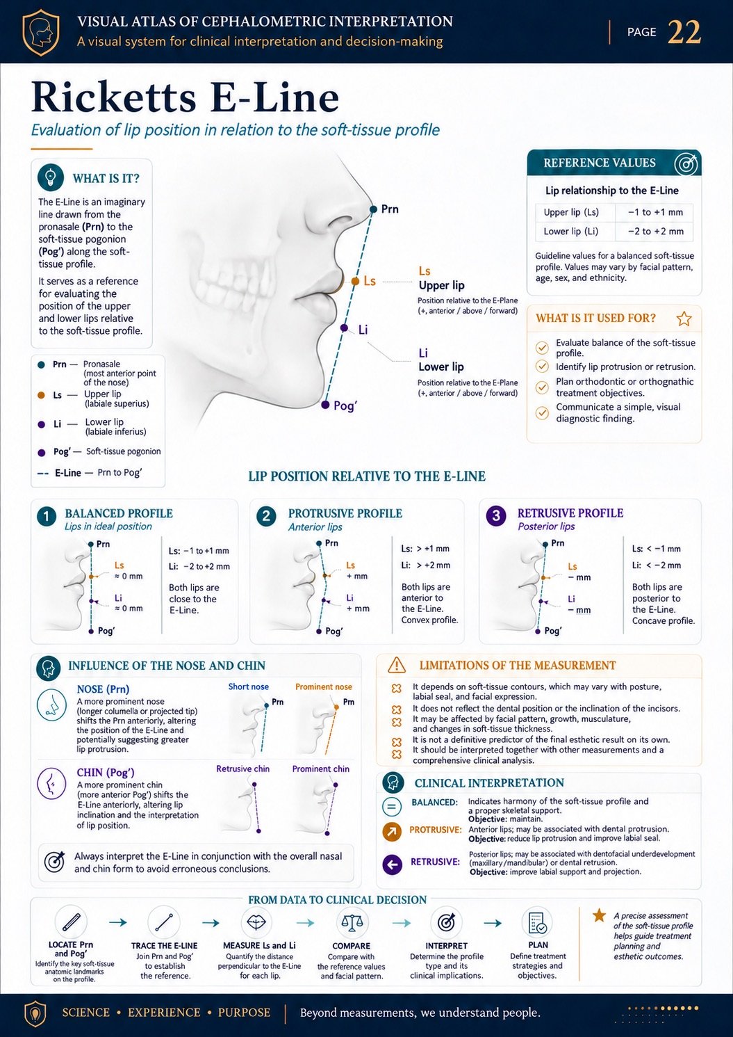
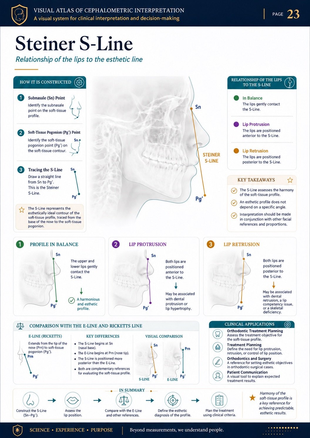
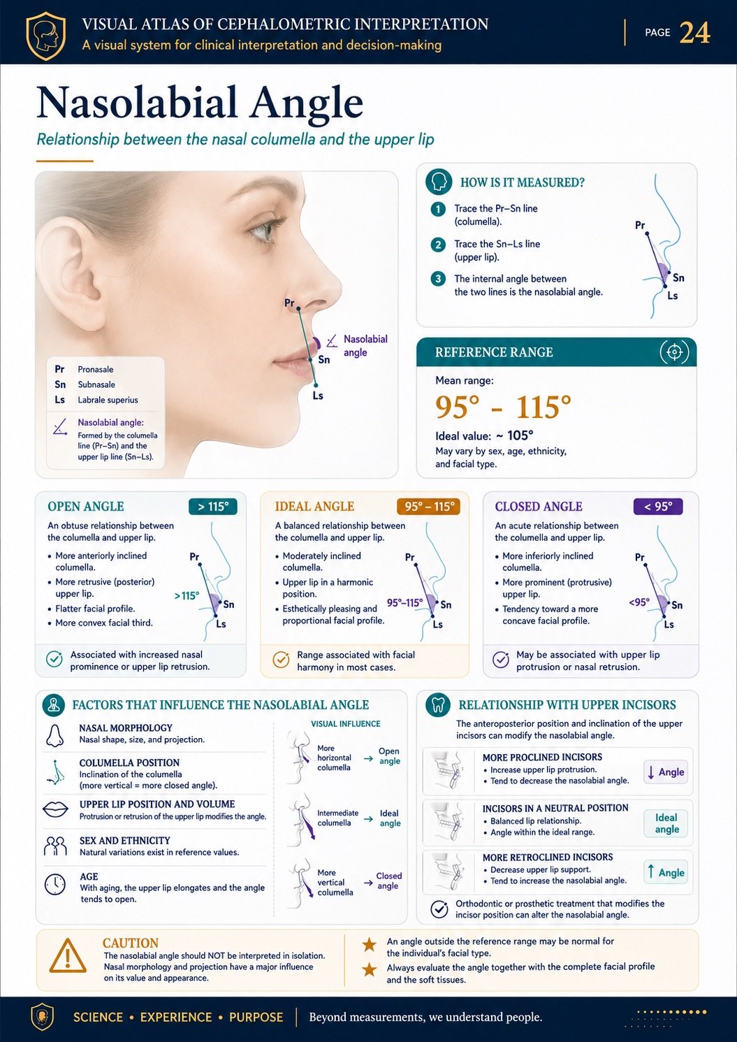
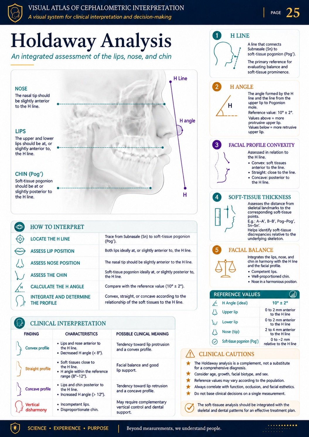
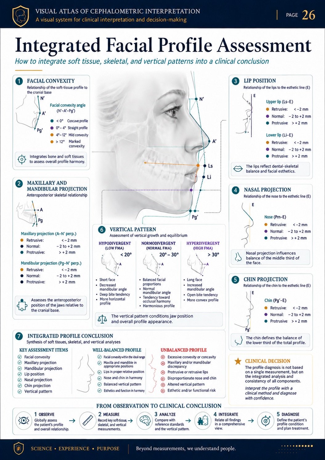
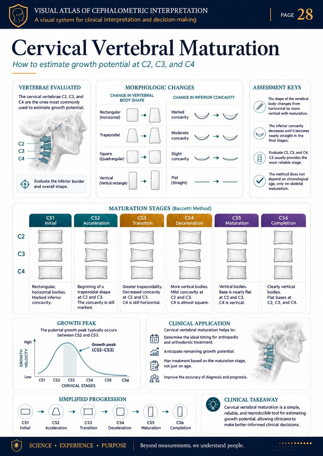
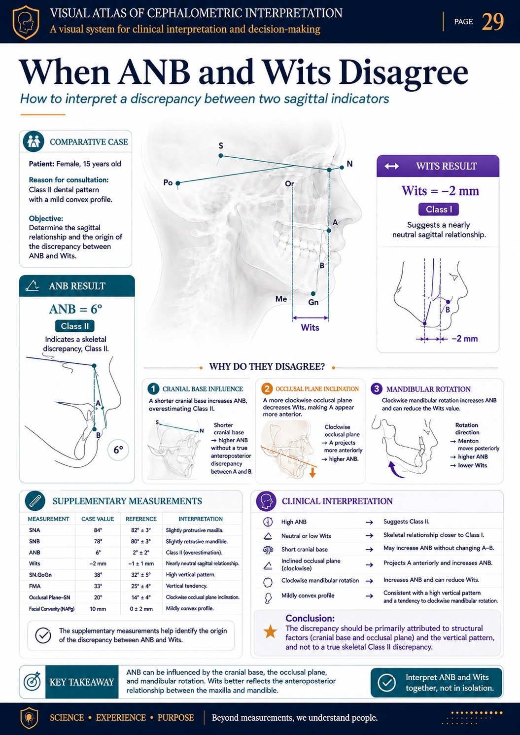
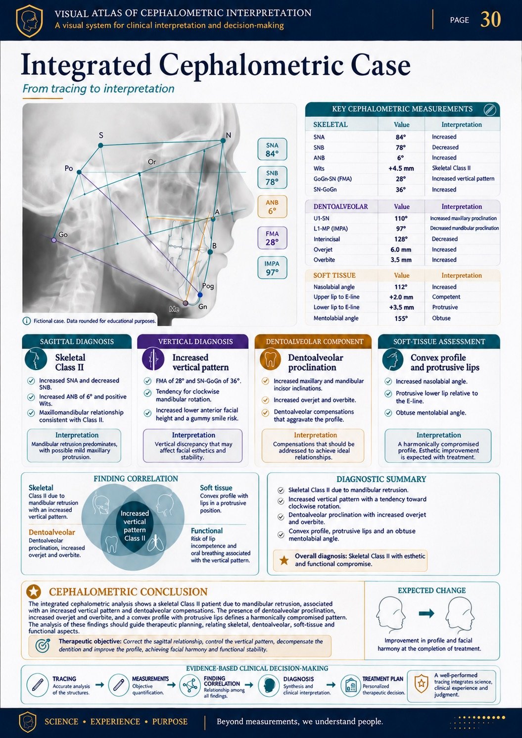
12 PDFs for understanding cephalometrics visually
Organized content to help you study, interpret, and reference the main components of cephalometric analysis.
Cephalometric Foundations
10 cards covering lateral cephalograms, image quality, measurement types, reference values, limitations, and interpretation workflow.
Anatomy, Cephalometric Landmarks, and Planes
10 cards covering skeletal, dental, and soft-tissue landmarks, planes, and reference lines.
Sagittal Assessment
10 cards covering SNA, SNB, ANB, Wits, convexity, and maxillomandibular relationships.
Vertical Assessment
10 cards covering FMA, SN-GoGn, the Y-axis, facial heights, divergence, and mandibular rotation.
Dentoalveolar Component
10 cards covering IMPA, 1-NA, 1-NB, the interincisal angle, inclination, protrusion, and dental compensation.
Soft Tissues and Facial Profile
10 cards covering the E-line, S-line, Holdaway analysis, the nasolabial angle, lips, nose, chin, and facial balance.
Steiner and Wits Analyses
10 cards explaining their key measurements, relationships, applications, and limitations.
Ricketts Analysis
10 cards covering fields, factors, axes, dental relationships, facial pattern, and soft tissues.
McNamara, Tweed, Downs, and Jarabak
10 comparison cards covering the main measurements and applications of each analysis.
Growth and Maturation
10 cards covering growth direction, facial patterns, cervical vertebral maturation, and growth potential.
Discrepancies and Integrated Cases
10 cards with divergent results, measurement correlation, and complete cephalometric cases.
Digital Workflow, AI, and Cephalometric Reports
10 cards covering automated tracing review, common errors, findings integration, and report preparation.
Explore the Visual System
Illustrated cards, tracings, comparisons, visual maps, and organized cases designed to simplify interpretation.
Who is the Visual Atlas for?
The Visual Atlas is designed for anyone who wants a more visual, organized approach to understanding and interpreting cephalometrics.
- Dental students who want to understand cephalometric foundations
- Orthodontic residents and graduate students
- Orthodontists seeking a quick visual reference
- Professionals who find it difficult to integrate measurements and findings
- Anyone reviewing landmarks, planes, angles, and reference values
- Professionals who use digital software and need to review result interpretation
Limited-time offer
Get the complete Visual System for Cephalometric Interpretation, including all 12 core PDFs and every bonus shown on this page.
Get instant access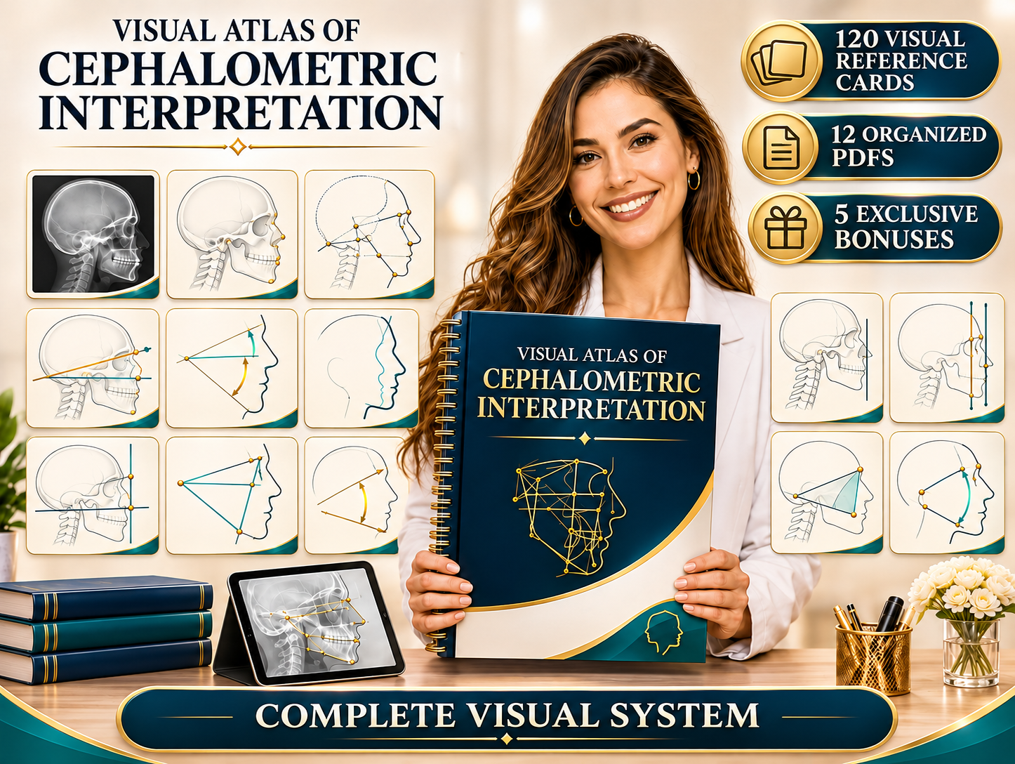
Everything included in the Visual System
Everything is organized so you can study each component progressively and reference the material whenever you need it.
- Cephalometric Foundations
- Lateral cephalogram
- Image quality control
- Skeletal cephalometric landmarks
- Dental and soft-tissue landmarks
- Reference planes and lines
- Linear and angular measurements
- Sagittal assessment
- Vertical assessment
- Skeletal Classes I, II, and III
- Facial growth pattern
- Incisor position and inclination
- Dentoalveolar compensation
- Soft tissues and facial profile
- Steiner analysis
- Ricketts Analysis
- McNamara, Tweed, Downs, and Jarabak
- Growth and cervical vertebral maturation
- Discrepancies between measurements
- Integrated cephalometric cases
- Digital tracing review
- Common interpretation errors
- Cephalometric report template
- C.L.A.V.E. interpretation method
Bonuses included with your access
Supplementary resources to simplify interpretation, review, and report preparation.
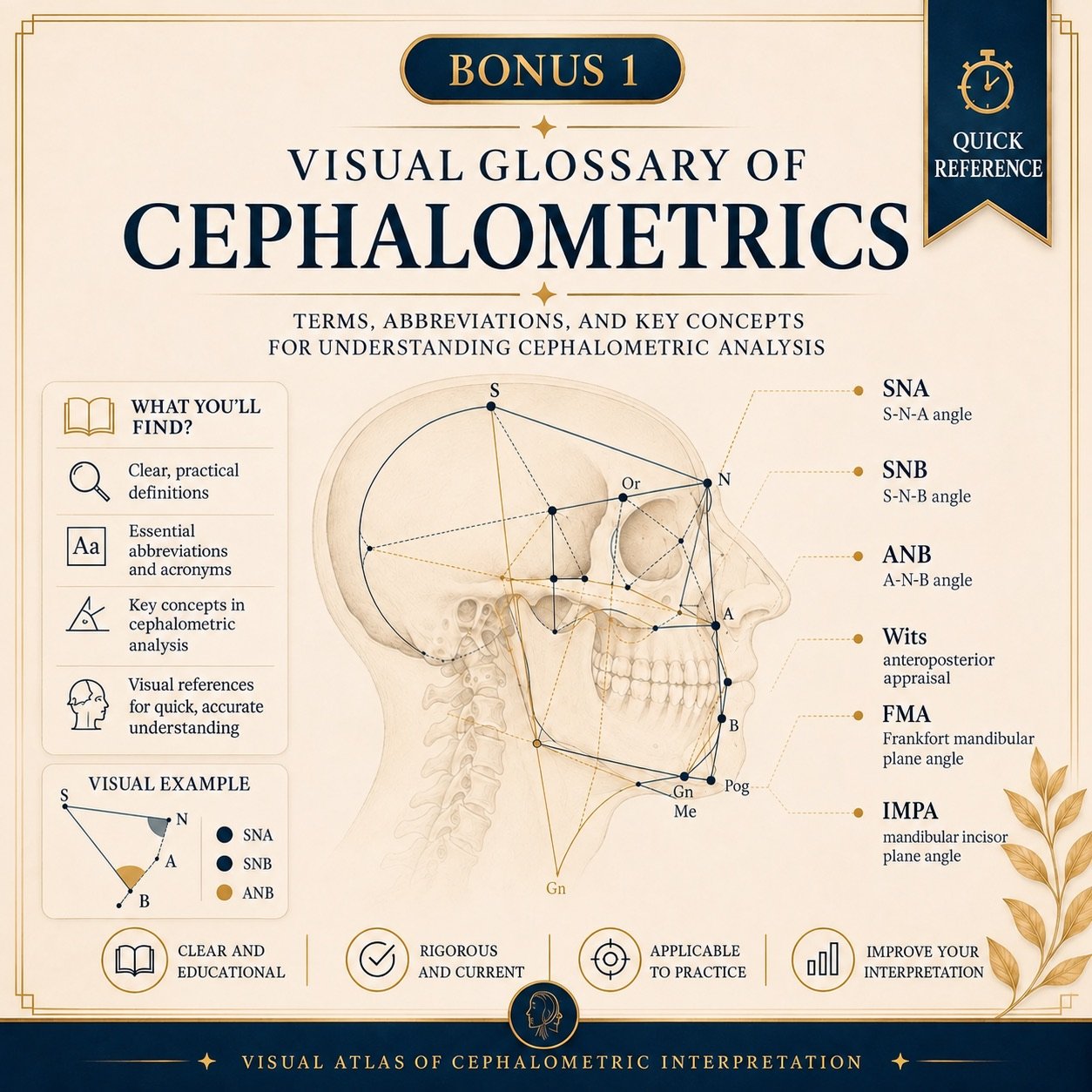
Visual Cephalometrics Glossary
Key terms, abbreviations, and concepts (SNA, SNB, ANB, Wits, FMA, IMPA) with clear definitions and visual references.
Understand cephalometric terminology without searching multiple sources.
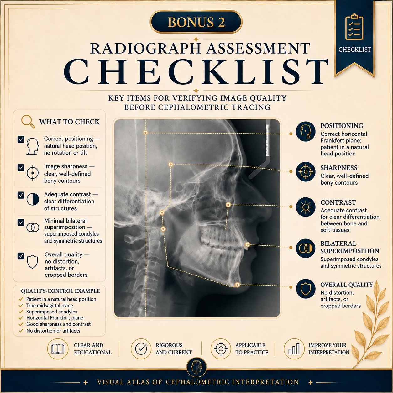
Radiograph Evaluation Checklist
Key checks for positioning, sharpness, contrast, and bilateral superimposition before tracing.
Begin every analysis with a reliable radiograph.
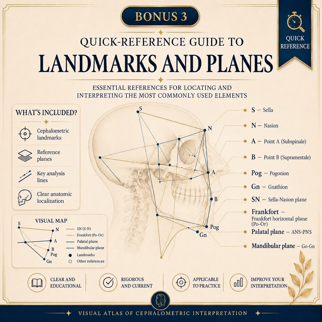
Quick Guide to Landmarks and Planes
Essential references for locating S, N, A, B, Pog, and Gn, plus the SN, Frankfort horizontal, palatal, and mandibular planes.
Locate every reference with anatomical clarity.
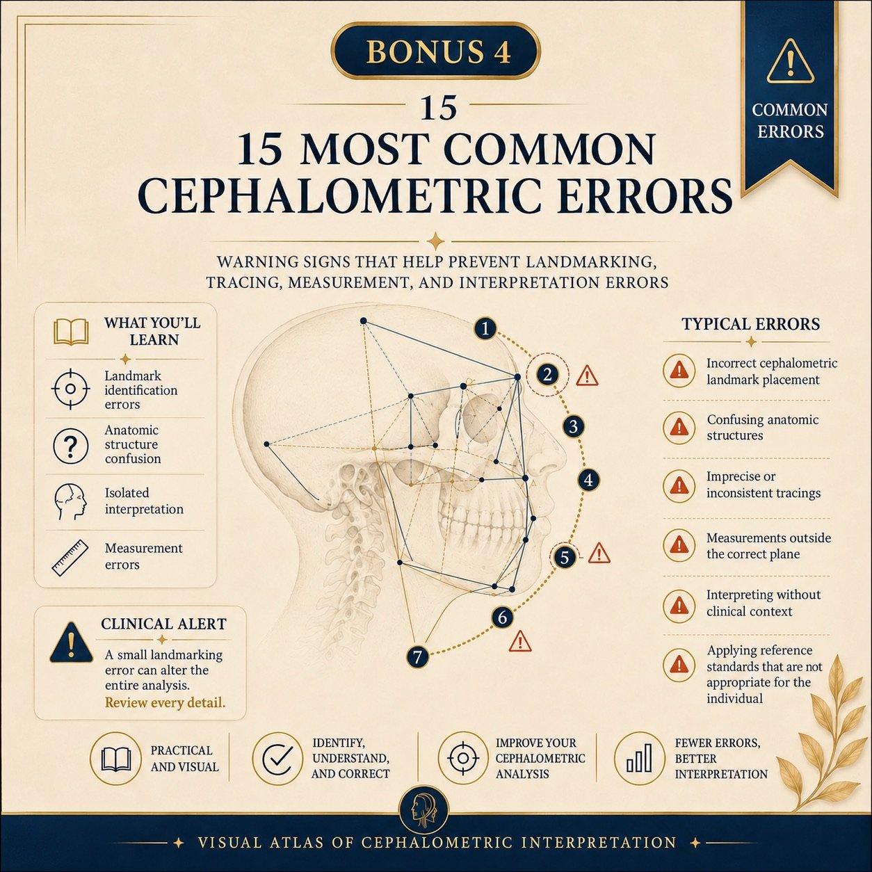
The 15 Most Common Cephalometric Errors
Warning signs that help prevent landmarking errors, structural confusion, off-plane measurements, and isolated interpretation.
Fewer errors, better clinical decisions.

7-Day Study Plan
A day-by-day visual plan: radiographs and landmarks, planes, sagittal and vertical diagnosis, dentoalveolar analysis, soft tissues, and integration.
Move forward step by step with a clear path.
Get access in minutes
See how simple it is to get started.
Complete your order
Choose your plan and check out securely.
Check your email
We will email your access instructions as soon as payment is confirmed.
Download the PDFs
Download the files directly to your device.
Study anywhere
Open the material on your phone, tablet, or computer.
Reference it anytime
Review the visual cards whenever you need them.
Students and dental professionals using the Visual Atlas
See how students and dental professionals are using a more visual approach to cephalometrics.
I always knew how to take the measurements, but I did not know what to do with them. The sagittal analysis cards finally helped me move from a number to a clinical conclusion.
The visual glossary and the guide to landmarks and planes made a huge difference in my seminars. I can locate Nasion, Sella, and Gonion without hesitation and no longer confuse the reference planes.
The vertical growth and mandibular rotation cards explain on one page what used to take me weeks to organize. The pattern comparison is exceptionally clear.
I use the radiograph evaluation checklist before every tracing. It has significantly reduced image-quality and structure-identification errors.
The Ricketts E-line card and Holdaway analysis gave me concrete language for explaining the profile to patients. The consultation became much more visual.
The 15 most common errors bonus reflected everything I had been doing wrong. I corrected my landmarking, and my measurements became reproducible.
I followed the 7-day study plan and arrived at the exam with the concepts organized: skeletal classes, vertical pattern, and dental analysis, each in its proper place.
Having the 12 PDFs organized by topic changed my routine. I print the card I need, keep it next to the radiograph, and reach a diagnosis much faster.
I always knew how to take the measurements, but I did not know what to do with them. The sagittal analysis cards finally helped me move from a number to a clinical conclusion.
The visual glossary and the guide to landmarks and planes made a huge difference in my seminars. I can locate Nasion, Sella, and Gonion without hesitation and no longer confuse the reference planes.
The vertical growth and mandibular rotation cards explain on one page what used to take me weeks to organize. The pattern comparison is exceptionally clear.
I use the radiograph evaluation checklist before every tracing. It has significantly reduced image-quality and structure-identification errors.
The Ricketts E-line card and Holdaway analysis gave me concrete language for explaining the profile to patients. The consultation became much more visual.
The 15 most common errors bonus reflected everything I had been doing wrong. I corrected my landmarking, and my measurements became reproducible.
I followed the 7-day study plan and arrived at the exam with the concepts organized: skeletal classes, vertical pattern, and dental analysis, each in its proper place.
Having the 12 PDFs organized by topic changed my routine. I print the card I need, keep it next to the radiograph, and reach a diagnosis much faster.
Start your visual cephalometrics study today
Get the complete Visual System, all 12 PDFs, and the visual tools you need to understand every measurement in context.
Start understanding cephalometrics
Essential version: compare it with the Complete plan to see the additional resources.
- Complete Visual System for Cephalometric Interpretation
- 120 visual reference cards
- 12 PDFs organized by topic
- Digital PDF files
- Instant access after payment
- Lifetime access
- Compatible with phones, tablets, and computers
- One-time payment
Visual Atlas + 5 Exclusive Bonuses
- 120 visual reference cards for Cephalometric Interpretation
- 12 complete, organized PDFs
- 5 exclusive bonuses to accelerate your understanding
- Visual Table of Reference Values and Ranges
- 7-Step Cephalometric Checklist
- Cervical Vertebral Maturation Guide
- Tracing Error Atlas
- Editable Report Template
- Digital material with instant access
- Lifetime access
- Future updates to this edition
- One-time payment

Your purchase is protected
You will have 7 days from purchase confirmation to review the material. If the product does not match what is presented on this page, you may request a refund within that period, subject to the terms shown at checkout.
Frequently asked questions
Get answers to the most common questions before purchasing.
The material is 100% digital and delivered as PDF files. No physical product is shipped.
After payment is confirmed, access instructions will be sent to the email address used at checkout.
Access is lifetime, subject to the delivery platform's terms.
No. You can study directly on your phone, tablet, or computer. Printing is optional and for individual use only.
It is designed for dental students, residents, graduate students, orthodontists, and professionals who want to review cephalometric interpretation visually.
No. The Visual Atlas of Cephalometric Interpretation is an educational, supplementary resource. It does not replace academic training, clinical evaluation, additional examinations, professional supervision, or an orthodontist's clinical judgment.
No. The material presents classic values and reference ranges, but results may vary by age, sex, individual characteristics, study population, and method. No measurement should be interpreted in isolation.
Yes. The material can support review of landmarks, measurements, and relationships generated by digital software. The professional remains responsible for verifying the tracing and completing the interpretation.
No. Your purchase grants an individual-use license. Sharing, copying, distributing, reselling, or publishing the files is prohibited.
The complete product and its supporting resources are provided in English.